AI in PET and Nuclear Medicine Imaging: Revolutionizing Molecular Diagnostics
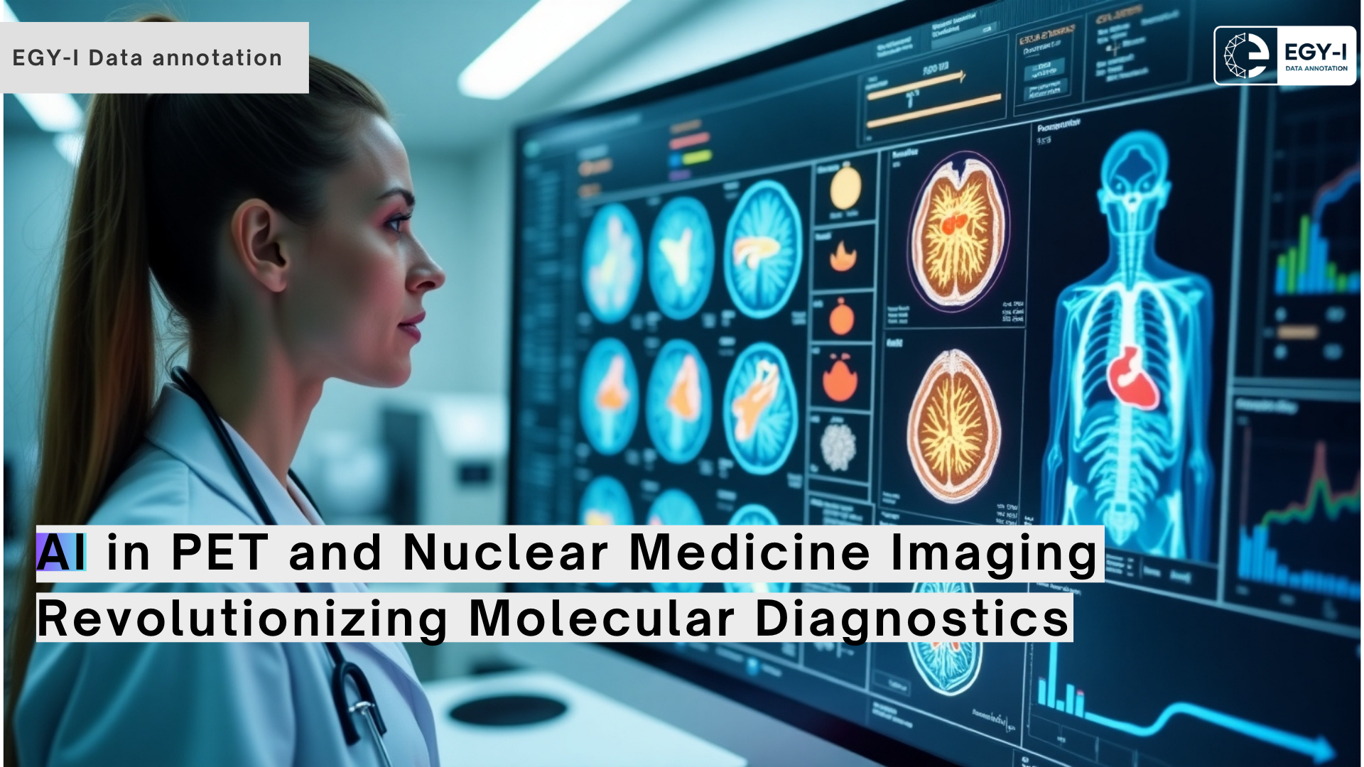
AI in PET and Nuclear Medicine Imaging: Revolutionizing Molecular DiagnosticsPositron Emission Tomography (PET) and other nuclear medicine imaging modalities are at the forefront of personalized medicine, enabling molecular-level visualization of physiological and pathological processes. From oncology to cardiology and neurology, these techniques offer unparalleled insights into disease mechanisms, guiding diagnosis, treatment planning, and monitoring. However, the growing demand for precision, coupled with the complexity of nuclear imaging data, poses significant challenges for healthcare providers.Enter Artificial Intelligence (AI)—a game-changer in the realm of PET and nuclear medicine imaging. AI technologies are transforming the way these modalities are utilized, improving diagnostic accuracy, streamlining workflows, and unlocking the potential for novel clinical applications. At EGY-I, we specialize in annotation services that form the foundation for AI development in nuclear medicine, supporting innovation in this rapidly evolving field.
Challenges in PET and Nuclear Medicine Imaging
- Complex Data Analysis: PET and nuclear medicine scans generate intricate datasets that require interpretation of dynamic biological processes.
- Noise and Artifacts: Imaging data often contains noise and artifacts that can obscure important findings.
- High Demand for Expertise: Accurate interpretation requires skilled professionals, and there is a global shortage of nuclear medicine specialists.
- Quantitative Analysis: Measuring tracer uptake values (e.g., SUV) for diagnosis and treatment response assessment is time-consuming and prone to variability.
AI addresses these challenges by automating processes, enhancing image quality, and enabling more consistent and accurate quantitative analysis.
AI Applications in PET and Nuclear Medicine Imaging1. Image Reconstruction and EnhancementAI-driven algorithms improve the quality of PET and nuclear medicine images:
- Noise Reduction: AI reduces noise in images, enabling lower-dose scans without compromising diagnostic accuracy.
- Faster Reconstruction: Deep learning accelerates image reconstruction, reducing scan times and enhancing patient comfort.
- Improved Resolution: Super-resolution techniques enhance the clarity of images, aiding in the detection of small lesions.
2. Tumor Detection and SegmentationIn oncology, AI excels in identifying and segmenting tumors:
- Automated Detection: AI identifies lesions with high sensitivity, even in complex cases.
- Tumor Volume Segmentation: Algorithms calculate the size and volume of tumors, aiding in treatment planning and monitoring.
- Tracer Uptake Analysis: AI measures standardized uptake values (SUVs) to evaluate metabolic activity and treatment response.
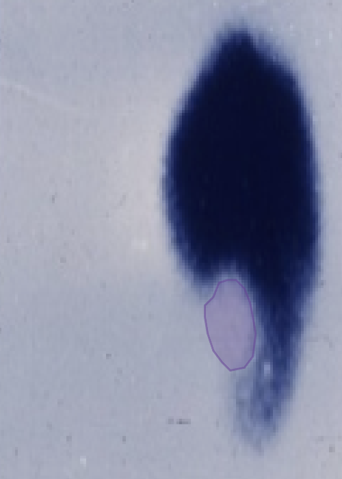 Liver solitary focal lesion
3. Cardiac ApplicationsAI enhances the diagnostic capabilities of nuclear cardiology:
- Perfusion Analysis: Automated detection of perfusion defects improves the accuracy of myocardial ischemia diagnosis.
- Ejection Fraction Calculation: AI estimates left ventricular ejection fraction from gated PET scans, streamlining functional assessment.
- Quantitative Flow Reserve: AI facilitates advanced metrics like myocardial blood flow, providing deeper insights into cardiac health.
4. Neurological ApplicationsIn neurology, AI is driving advancements in imaging for disorders like Alzheimer’s and epilepsy:
- Early Alzheimer’s Detection: AI identifies amyloid plaques and other biomarkers in PET scans, enabling early diagnosis and intervention.
- Seizure Focus Localization: AI enhances the accuracy of detecting epileptogenic zones in brain imaging.
- Neuroinflammation Analysis: Algorithms quantify tracer uptake associated with inflammation, aiding in research and diagnosis.
5. Workflow AutomationAI streamlines nuclear medicine workflows:
- Report Generation: Automated tools generate preliminary findings, saving time for nuclear medicine specialists.
- Case Prioritization: Algorithms prioritize critical cases for review, ensuring timely intervention.
The Role of Image Annotation in AI for PET and Nuclear MedicineThe development of AI models for PET and nuclear medicine imaging relies on meticulously annotated datasets. At EGY-I, we provide specialized annotation services to support these efforts.Our Annotation Capabilities:
- Lesion Annotation: Accurate labeling of tumors, metastases, and other abnormalities.
- Organ Segmentation: Detailed segmentation of organs such as the brain, heart, and liver.
- Tracer Uptake Mapping: Annotation of regions with abnormal tracer activity.
- Artifact Identification: Highlighting noise and artifacts for algorithm training.
Our expertise ensures that your AI solutions are built on high-quality, reliable datasets.
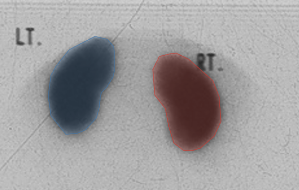
Kidney segmentation
How EGY-I Supports AI in Nuclear MedicineAt EGY-I, we empower AI innovation in PET and nuclear medicine imaging through our comprehensive annotation services.Why Choose EGY-I?
- Experienced Team: Our annotators are well-versed in the intricacies of nuclear medicine imaging.
- Customized Solutions: We adapt to your project needs, delivering annotations tailored to specific AI applications.
- Quality Assurance: Rigorous quality checks ensure accurate and consistent annotations.
- Timely Delivery: We prioritize meeting deadlines without compromising on quality.

AI is revolutionizing PET and nuclear medicine imaging, enabling earlier diagnoses, personalized treatments, and more efficient workflows. At EGY-I, we are proud to contribute to this transformation by providing the high-quality annotated datasets that AI models need to succeed.
